
Pemphigus Foliaceus in Dogs: Symptoms, UV Triggers, and Nose Protection
TLDR
Pemphigus foliaceus (PF) is the most common autoimmune skin disease in dogs. Like Discoid Lupus Erythematosus (DLE), it causes crusting and sores on the nose and face, and UV light is a documented trigger that makes it worse. PF requires veterinary diagnosis and immunosuppressive treatment, but consistent sun management - including a physical nose barrier - is part of the protocol and something owners control every day.
If your dog has recurring crusty sores on the nose, face, or ears that flare up in summer and ease off in winter, pemphigus foliaceus may be what you are dealing with.
What Is Pemphigus Foliaceus?
In a healthy dog, skin cells are held together by adhesion proteins. In PF, the immune system produces antibodies that attack one of these proteins - desmoglein 1 - causing the outer skin layers to separate. The result is blistering, pustules, and the characteristic crusty lesions that owners first notice on the face and nose.
PF affects only the outermost skin layer, which is why lesions tend to be superficial but recurring and widespread. Unlike some deeper autoimmune conditions, PF rarely involves internal organs, but the skin involvement can be severe enough to affect a dog's appetite, energy, and quality of life.
Symptoms: What to Look For
PF lesions follow a recognizable pattern. They typically start on the face and are often symmetrical, appearing on both sides of the body at once.
Nose and nasal planum: Crusty, scabbed lesions on the hairless part of the nose. The surface may lose its normal cobblestone texture and become raw or depigmented.
Face and muzzle: Pustules and erosions on the dorsal muzzle, often spreading toward the eyes and forehead.
Ears: The ear flaps are frequently involved, sometimes before the nose shows any symptoms.
Paw pads: A distinguishing feature of PF - the footpads often develop lesions and crusting alongside the face. Bacterial pyoderma, which looks similar, rarely affects both face and paw pads at the same time.
General signs in severe cases: Lethargy, reduced appetite, fever, and noticeable itching.

Why Summer Makes It Worse
One of the most consistent patterns in PF is its seasonal nature. Lesions tend to worsen in summer and improve in winter. This directly reflects the role of UV light as a trigger.
Veterinary research has documented that exposing dogs with facial PF to UVB radiation increases the breakdown of the skin cell adhesion that holds tissue together - the exact mechanism that drives the disease. Sun management is not a supplementary tip. It is part of the treatment protocol, alongside medication.
Which Breeds Are Most at Risk?
While any dog can develop PF, certain breeds appear significantly more often in clinical studies:
Akita and Chow Chow - most consistently overrepresented in PF studies
Bearded Collie, Newfoundland, Schipperke
Doberman Pinscher, Cocker Spaniel, Dachshund, Shar-Pei, Collie
PF most commonly develops in middle-aged dogs, around 5 years old on average, though it can appear at any age.
PF vs. DLE: What Is the Difference?
Pemphigus foliaceus and Discoid Lupus Erythematosus are the two most common autoimmune skin diseases in dogs. Both affect the nose and face, both are worsened by UV, and both require long-term management. Owners researching one often have a dog with the other.
Location: DLE is almost exclusively confined to the nose. PF also affects ears, paw pads, and can spread to the trunk.
Severity: PF is generally more aggressive. Systemic symptoms like fever and lethargy are possible with PF and rare with DLE.
Treatment: DLE can often be managed with topical medication and UV avoidance. PF typically requires systemic immunosuppression.
Paw involvement: Footpad lesions strongly point to PF, not DLE.
UV connection: Both conditions are UV-triggered and UV-worsened. Sun management helps both.
There is also a crossover form - pemphigus erythematosus - which shares features of both PF and DLE, primarily affecting the nose and face and especially sensitive to sunlight.
Diagnosis
PF looks similar to bacterial pyoderma and cannot be diagnosed from appearance alone. Misdiagnosis changes the entire treatment approach. Veterinary diagnosis typically involves:
Skin biopsy: The definitive test. A tissue sample is sent to a veterinary dermatopathologist who looks for acantholytic cells - loose keratinocytes that have separated from surrounding skin.
Cytology: A quicker in-clinic option. Material from an intact pustule is examined under a microscope for characteristic cells alongside neutrophils.
Blood work: CBC and biochemistry panel to rule out other causes and establish a baseline before starting immunosuppressive therapy.
Treatment
There is no cure for pemphigus foliaceus. The goal of treatment is remission - reducing active lesions and keeping the immune response suppressed enough for the skin to heal. Standard treatment typically combines:
Corticosteroids (prednisone/prednisolone) as the first-line medication, starting at higher doses to achieve remission, then gradually tapered
Steroid-sparing immunosuppressants such as cyclosporine or azathioprine, often added to reduce long-term steroid side effects
Topical therapy for milder or localized cases - tacrolimus or steroid creams used alongside or instead of systemic medication
Antibiotics when secondary bacterial infections are present, which is common
Sun avoidance consistently recommended by veterinary dermatologists as an essential part of management - not optional
Approximately 50% of dogs with PF achieve complete remission with treatment, and around 35% achieve partial remission. Relapses are common and many dogs require lifelong maintenance medication. Regular vet monitoring every 6 months is important for dogs on long-term immunosuppressants.
UV Protection: The Part Owners Manage Daily
Medication manages the immune response. UV management manages the trigger. For PF dogs with nose and facial lesions, consistent protection reduces flare frequency - particularly during summer when UV index is highest.
Walk your dog early morning or evening, outside peak UV hours (10am to 4pm)
Limit time on reflective surfaces like concrete and water in direct sunlight
Use a dog-safe sunscreen on exposed areas - avoid zinc oxide, which is toxic if ingested. See our guide to sunscreen safety for dogs.
Use a physical barrier over the nasal planum when sun exposure is unavoidable
A 2023 peer-reviewed study in Veterinary Dermatology (Milich et al.) tested physical UV nose guards on dogs and found they blocked up to 99% of ultraviolet radiation, with statistically significant results (p<0.001). Physical barriers solve the reapplication problem that makes sunscreen impractical for most dogs. Read the study on PubMed →
How SnoutCover Fits Into PF Management
SnoutCover was developed for dogs with DLE, but the problem it solves is identical for PF dogs with nasal and muzzle lesions. The nose is exposed to UV, topical medications get licked off immediately, and there is no practical way to maintain protection through the day.
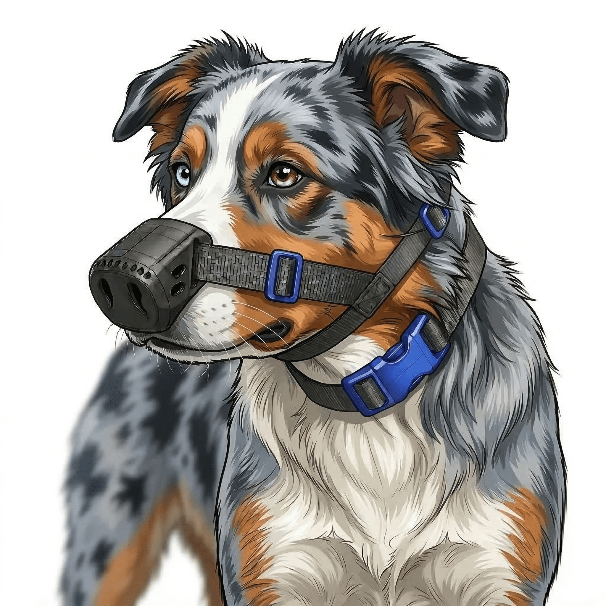
SnoutCover is a 3D-printed TPU nose shield that blocks UV from reaching the nasal planum, holds topical medication in place so it absorbs instead of being licked away, and allows normal eating, drinking, and breathing through a ventilated design.
It does not replace veterinary treatment. PF requires medication, monitoring, and regular vet care. But for the UV management piece - the part of the protocol owners handle daily - SnoutCover provides consistent, hands-free protection that sunscreen alone cannot match.
SnoutCover protects and supports - it does not replace your vet
SnoutCover blocks UV and keeps treatments in place. It is not a cure and not a wound dressing. If your dog has lesions on the nose, face, ears, or paws, see your veterinarian first to get a definitive diagnosis and treatment plan before adding any physical barrier to the routine.
Frequently Asked Questions
Is pemphigus foliaceus the same as DLE in dogs?
No, but they are related. Both are autoimmune skin diseases that affect the nose and face and are worsened by UV. DLE stays confined to the nose; PF also affects ears, paw pads, and sometimes the trunk, and it tends to be more aggressive.
Which dog breeds are most prone to pemphigus foliaceus?
Akitas and Chow Chows are the most consistently overrepresented breeds. Bearded Collies, Newfoundlands, Doberman Pinschers, Cocker Spaniels, and Dachshunds are also at higher risk.
Can UV light trigger pemphigus foliaceus in dogs?
Yes. UV exposure is a documented trigger and worsens active lesions. Seasonal flares - worse in summer, better in winter - are one of the hallmark patterns of PF. Sun management is part of the veterinary treatment protocol.
How do I protect my dog's nose with pemphigus foliaceus?
Avoid peak-sun walks, use a dog-safe sunscreen without zinc oxide, and add a physical UV barrier to keep protection in place through the day. See our cream-plus-cover guide for the combined approach.
Can pemphigus foliaceus in dogs be cured?
No. PF is managed, not cured. With vet-directed immunosuppressive treatment and consistent UV protection, approximately 50% of dogs achieve complete remission. Many dogs need lifelong maintenance therapy and regular monitoring.
